

Cutting-edge veterinary equipment elevates Cornell’s patient care

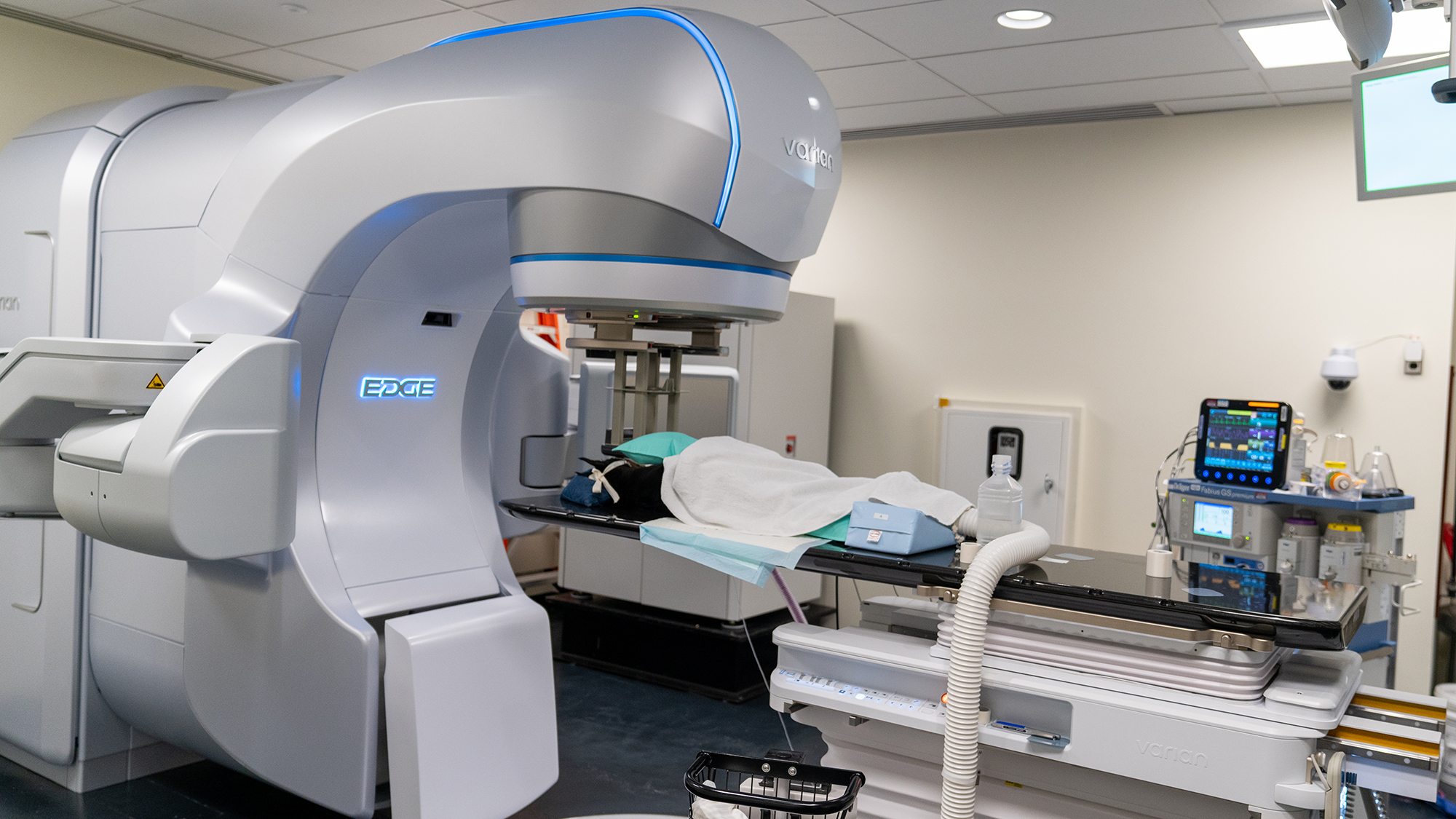
The Varian Edge linear accelerator provides state-of-the art, non-invasive radiotherapy treatments. Photo: Carol Jennings/CVM
It sounds like futuristic equipment only seen in science fiction — medical machines capable of targeted radiation that avoids healthy tissues, scanners that count the energy in photons to create three-dimensional images.
Such machines are now a reality at the Cornell University Hospital for Animals, which has adopted the Varian Edge linear accelerator and Siemens Naeotom Alpha photon-counting CT scanner, the most advanced technology of their kind on the market today, enhancing both therapy and diagnosis for patients.
“It’s important that Cornell stays on the cutting edge of what veterinary medicine has to offer. Installing crucial pieces of technology allow us to increase our patient care offerings and advance the field through research and teaching,” said Susan Ruland, interim hospital director.
The Varian Edge linear accelerator provides state-of-the art, non-invasive radiotherapy treatments. The closest comparable facilities are in New York City, and while approximately 40% of veterinary schools have radiation oncology programs, few have access to such technology. Cornell’s Varian Edge replaces machinery previously installed in 2000.

“The technology advances every 10 to 15 years. It’s important to keep the service progressing alongside the technology,” said Dr. Tracy Gieger, clinical professor in the Section of Radiation Oncology. Gieger was also a resident at the hospital from 2005-2007 and worked with the previous equipment.
“You’d be hard-pressed to find technology in any veterinary center in any part of the world that can match our linear accelerator for versatility, spatial resolution or safety standards,” said Dr. Parminder Basran, associate research professor in the Section of Medical Oncology. Over the last two years, Basran led the team that selected the linear accelerator and oversaw its installation.
Speed, precision
A key component of the new linear accelerator is how it fast-tracks treatment times. In the past, for example, a patient with a brain tumor might need to come in 20 times for radiation therapy treatments. Now, such treatments are possible within five visits, usually over the course of a week. This reduces the amount of time an animal spends under anesthesia, limits the commuting time by an owner who may be out of state — and importantly opens up more treatment times for other patients.
The new linear accelerator is also more precise than older versions. “We can essentially sculpt a dose of radiation around normal tissues,” Gieger said. For example, if a dog has a tumor next to its eye, previous generations of the technology weren’t necessarily able to shield the eye from radiation during treatment. “Basically, the eye adjacent to the tumor would be getting 90% of the dose as well — now, it’s down to 10 or 20%.” This precision not only protects the eye, but decreases other side-effects from radiation treatments more generally.
Although radiotherapy treatments are a primary function of the linear accelerator, it has the capabilities to do much more. “This new system has an abundance of features that open up so many possibilities,” Basran said.
Indeed, the hardware was upgraded, but the software supporting that hardware has gone through evolutions, too. “In the past, we might not have been as concerned about information databases, but it’s critical in today’s world,” Basran said. “So not only can we deliver exquisite forms of radiation therapy, but we can manage the records, imaging data and metadata associated with these patients and more. It’s a very robust information system.”
Better image quality
The linear accelerator is one of several recent upgrades at the Cornell University Hospital for Animals — among which is a new Siemens Naeotom Alpha photon-counting CT scanner. This machine is a leap in efficient imaging capabilities, with increased sensitivity, reduced radiation exposure and more powerful detection. It is similarly replacing technology from around 2000.
“Photon-counting CT is the newest CT technology, with fewer than 50 scanners in clinical use, and this is the first installed in veterinary medicine,” said Peter Scrivani ’89, D.V.M. ’93, professor and section chief of imaging.
Dr. Ian Porter, assistant clinical professor in the Section of Diagnostic Imaging, along with the other imaging team members, searched for new equipment that would enhance their clinical abilities, facilitate cutting edge research and allow the hospital’s imaging technology to stay up to date for many years to come.
“A CT scan is an integral part of case management for many patients, including emergency trauma patients, in complicated vascular malformations, in cancer patients for surgical and radiation planning, as well as staging to help identify the potential spread of a tumor, in orthopedic surgery and sports medicine, in exotic animal medicine, and in large animal medicine and surgery cases,” Porter said. “We were looking to adopt a technology that sets the bar in imaging and allows us to provide the best care to our patients.”
A photon-counting CT functions differently than typical scanners because it can detect and count each individual x-ray photon, which is a tiny particle of electromagnetic energy. This produces images with finer details. Furthermore, the new scanner records each photon’s energy level. “Because it can distinguish between different x-ray energies, it can separate materials that look similar in regular CT scans — such as bone, soft tissue and contrast agents — without needing multiple scans or extra equipment,” Scrivani said.
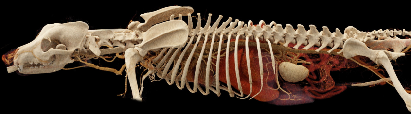
“It’s phenomenal the kind of detail you can see,” Porter said.
Moreover, the CT can work hand-in-hand with the linear accelerator, essentially creating a digital surrogate of the patient.
“We put it into our planning system and simulate what the linear accelerator’s radiation beams would look like,” Basran said. If a cat must have multiple treatments, experts can use advanced imaging from the new CT alongside the robotic couch of the linear accelerator to ensure the cat is positioned exactly as it was before. “This allows us the kind of precision that’s within half a millimeter, which is crucial when you’re dealing with small patients or tiny tumors.”
Dr. Rory Todhunter, orthopedic surgeon, professor of surgery and the inaugural director of the Cornell Richard P. Riney Canine Health Center, understands the impact that the new CT scanner can have on patient health at Cornell as well as on future research. “Supporting canine health research is a key pillar of the Riney Canine Health Center, and through our outreach efforts, the funds we raise are so important in supporting technology that fuels greater research. This is what will help our dogs live longer, healthier lives in the long run. We are pleased to be able to support such technology at Cornell.”
“The new CT scanner will benefit a wide range of clinical services throughout the hospital — including small animal, large animal and exotic species care,” Scrivani said. “It will be invaluable in diagnosing and managing conditions across all body systems and parts, including soft tissue surgical, medical, neurological, cardiovascular and orthopedic cases.”

Digital data collection, analysis
The linear accelerator and CT scanner will also support research at the College of Veterinary Medicine, including through translational medicine in oncology as well as non-oncologic uses like vision machine learning of medical images.
“It’s really starting to capture the imaginations of researchers, how we might be able to use this technology,” Basran said. For example, if a researcher wanted to examine patient toxicities from their treatments, in the past, they would review stacks of paper for that information and manually record them into a spreadsheet. “We have an opportunity to modernize that with digital templates to score toxicity,” he said. “Now we can query any patient, any time, any stage of treatment, see what the toxicity is, even pull that data and do retrospective analyses.”
As a medical physicist, Basran loves pushing the limits of technology. “It’s exciting,” he said. “This opens so many more opportunities for research, collaboration and opportunities to partner with scientists across campus and even globally.”
In addition to efficient treatment options, more precise therapies and increased research potential, the linear accelerator can address cardiac arrhythmias and osteoarthritis; it can block blood vessels around tumors, provide image-guided technology for specialty therapies, permit patient enrollment in certain clinical trials — and much more. The team is also exploring potential future uses in cardiology, small animal surgery, neurology and imaging settings.

Legacy of radiation oncology
Cornell has always been a leader in the veterinary radiation oncology specialty. Margaret McEntee, D.V.M. ’86, the C.V. Starr Professor of Medical and Radiation Oncology, is one of Cornell’s foundational oncologists and has trained many residents in the specialty over the years, including Gieger. With her retirement this spring, McEntee hands the baton to the next generation.
“She’s really grown the program, and we’re hoping to grow it even more thanks to the capabilities of the new technology,” Gieger said.
Choosing the right linear accelerator was no easy feat. In partnership with the Section of Medical Oncology, Basran chaired an investigation committee on the project, which launched in 2023. The committee researched, reviewed and examined the complicated variables involved in the selection process. In addition to technology, the committee evaluated personnel needs to support the upgrade. Every veterinary radiation facility has an affiliation with a medical physicist, but Basran is actually on-site at Cornell to work with the oncology team on radiation plans and for education purposes. The team has hired two additional radiation oncologists, and two radiation therapists who come from the human medicine realm.
Future trainees will have the opportunity to see Cornell’s upgraded equipment in action. “Residents will get to learn groundbreaking technologies in our training environment,” Basran said. “With an outstanding linear accelerator and CT scanner, it provides them fundamental knowledge while demonstrating the highest bar in patient care.”
This technology was made possible through the John and Eloise Turrel Endowment for Veterinary Oncology, the Richard P. Riney Canine Health Center, the Cornell Feline Health Center and donations from the Clays for K9 Cancer Research Benefit.

“This new equipment will have a three-fold positive impact on our operations: treating patients with the most effective and latest standard of care, teaching students and house officers, and providing cutting-edge technology that will foster the research that is essential to improving our ability to help our patients in the future,” said Dr. Bruce Kornreich, board-certified veterinary cardiologist in the Cornell University Hospital for Animals and director of the Cornell Feline Health Center (FHC). “These are all consistent with the college’s mission and why it was important for FHC to support this investment.”
The team is looking forward to leveraging the new technology not just for unique, rare cases, but for the broader population of clients. They’re also interested in pursuing a standing CT system that can accommodate patients over 300 lbs, which would advance imaging capabilities in the Cornell Equine and Nemo Farm Animal Hospitals and allow veterinarians to scan large animals without general anesthesia.
“Not only are we are committed to the highest standard of care for our patients, but investing in technology like this has far-reaching effects on recruitment and the future of profession as well,” said Ruland.
Written by Melanie Greaver Cordova


